
What Is Giemsa Stain?
Giemsa stain is a widely used laboratory staining technique that helps researchers, laboratory professionals, and medical specialists examine blood cells, parasites, chromosomes, and other biological samples under a microscope. It belongs to the Romanowsky family of stains and is particularly valuable because it provides clear differentiation between cellular structures.
The stain is named after Gustav Giemsa, a German chemist who developed the staining method in the early twentieth century. Since then, Giemsa staining has become an important technique in hematology, parasitology, cytogenetics, and microbiology.
One of the best-known applications of giemsa stain is the identification of malaria parasites in blood films. It can also help detect other blood parasites and provide useful information about the appearance and structure of different blood cells.
Because Giemsa stain produces contrasting colors in cellular components, it allows microscopic examination to be more informative than viewing an unstained specimen.
Why Is Giemsa Stain Important?
Microscopic specimens can contain many structures that are difficult to distinguish without staining. Giemsa stain improves visibility by interacting with different components of cells and microorganisms.
The technique is especially useful because it can highlight:
- Red blood cells
- White blood cells
- Platelets
- Cell nuclei
- Cytoplasmic structures
- Chromatin patterns
- Blood parasites
- Certain microorganisms
- Chromosomal regions
In diagnostic and research laboratories, the quality of staining can have a major influence on microscopic interpretation. A properly prepared Giemsa-stained slide can provide valuable morphological information that may otherwise be difficult to observe.
Giemsa Stain Composition
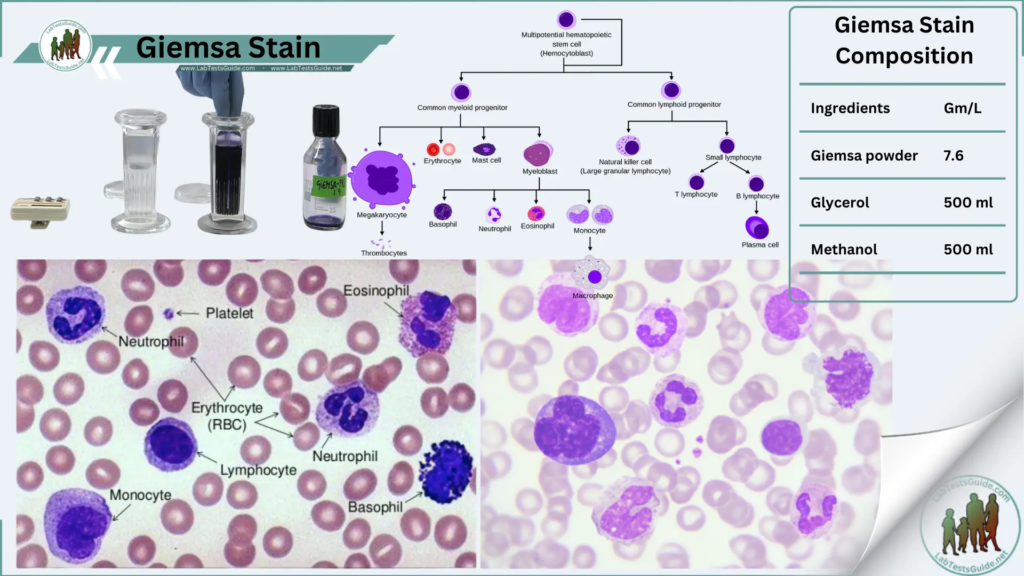
Giemsa stain is commonly prepared from a combination of dyes associated with the Romanowsky staining system. Its major staining components include azure dyes, methylene blue, and eosin.
These components interact differently with cellular structures.
Basic dyes generally bind more strongly to acidic cellular components, while acidic dyes interact with structures that have a more basic character. This difference produces the characteristic range of blue, purple, pink, and reddish shades seen on Giemsa-stained specimens.
The exact formulation and working concentration can vary depending on the manufacturer, laboratory protocol, and intended application.
Commercial Giemsa stains are often supplied as concentrated solutions that are diluted before use.
Principle of Giemsa Staining
The principle behind Giemsa staining is based on the interaction between dye components and different chemical structures within cells.
Nucleic acids and other acidic components tend to take up basic dye components, producing blue to purple coloration. Hemoglobin and some other cellular structures interact with eosin-containing components and may appear pink or reddish.
This differential staining creates contrast between various components of a specimen.
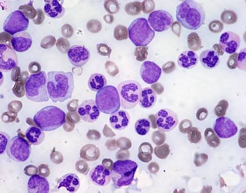
For example, in a peripheral blood film, red blood cells generally appear pale pink, while white blood cell nuclei can appear deep purple or blue. Parasites may display distinctive staining patterns that help laboratory professionals recognize their morphology.
The precise appearance depends on factors such as stain concentration, staining time, pH, specimen preparation, and slide quality.
Giemsa Stain for Blood Smears
One of the most important uses of Giemsa stain is the examination of peripheral blood smears.
A blood smear allows individual blood cells to be observed under a microscope. When properly stained, Giemsa provides useful information about cell size, shape, nuclear characteristics, cytoplasm, and intracellular structures.
The technique is commonly used in hematology laboratories for evaluating blood-cell morphology.
Giemsa staining can also be particularly useful when examining suspected blood parasites. Parasites may appear inside red blood cells or elsewhere in the blood film, depending on the organism and stage of development.
Giemsa Stain for Malaria Diagnosis
Giemsa stain has a long-standing role in malaria microscopy.
Malaria parasites belonging to the genus Plasmodium can be detected in stained blood films. Both thick and thin blood films may be used, with each preparation providing different advantages.
A thick blood film concentrates blood components and can improve the likelihood of detecting parasites when parasite levels are relatively low. A thin blood film preserves individual red blood cell morphology and can be useful for examining parasite characteristics.
Giemsa staining can help reveal different developmental stages of malaria parasites, including ring forms, trophozoites, schizonts, and gametocytes.
The microscopic appearance of these stages can provide important clues for parasite identification and laboratory diagnosis.
Thick Blood Film and Giemsa Stain
Thick blood films contain a larger volume of blood concentrated into a smaller microscopic area. During processing, red blood cells are largely disrupted, leaving white blood cells, platelets, and parasites more exposed.
This preparation can be particularly useful for detecting malaria parasites.
A properly stained thick film may allow the microscopist to identify parasites against a background containing stained cellular material.
However, interpretation requires appropriate training because the appearance of parasites in thick films differs from that in thin films.
Thin Blood Film and Giemsa Stain
Thin blood films are prepared by spreading a small amount of blood into a relatively thin layer across a microscope slide.
Unlike thick films, red blood cell morphology is preserved. This makes thin films particularly useful for studying parasite morphology and estimating the relationship between parasites and individual red blood cells.
Thin films can therefore provide additional information when identifying malaria species or evaluating blood-cell morphology.
In many laboratory settings, thick and thin films are used together because they complement one another.
Giemsa Stain Procedure
The exact Giemsa staining procedure varies between laboratories and applications, so the manufacturer’s instructions or validated laboratory protocol should always be followed.
A general blood-film staining workflow involves several stages.
1. Prepare the Blood Smear
A clean microscope slide is used to prepare either a thin or thick blood film. The specimen should be spread evenly and allowed to dry according to the applicable laboratory procedure.
2. Fix the Thin Film When Required
For many thin blood-film protocols, methanol fixation is used before staining. Thick blood films are generally handled differently because fixation can interfere with the intended preparation.
3. Prepare the Giemsa Working Solution
A concentrated Giemsa solution is diluted with an appropriate buffered solution. The correct dilution depends on the laboratory method and the purpose of staining.
4. Apply the Stain
The prepared Giemsa working solution is applied to the slide for the validated staining period.
5. Rinse Carefully
After staining, the slide is gently rinsed using the recommended buffered water or solution. Excessive washing can remove material or affect staining quality.
6. Dry the Slide
The stained slide is allowed to dry properly before microscopic examination.
7. Examine Under the Microscope
The slide is examined using appropriate microscopy techniques. Higher magnification, including oil immersion where applicable, may be used to assess cellular and parasitic structures.
Giemsa Stain Results
A good Giemsa stain produces clear contrast between different components of the specimen.
In a properly stained peripheral blood film:
- Red blood cells generally appear pale pink or salmon-colored.
- White blood cell nuclei commonly appear dark blue to purple.
- Cytoplasm may show varying shades of blue.
- Platelets may appear as small purple or violet structures.
- Nuclear chromatin can show strong purple coloration.
- Certain parasites may be visible with characteristic blue and purple staining patterns.
The final appearance can vary depending on the specimen, staining conditions, microscope, and preparation technique.
Giemsa Stain for Chromosomes
Giemsa staining also has an important role in cytogenetics.
A specialized technique known as G-banding uses Giemsa stain to produce characteristic banding patterns on chromosomes. These patterns help researchers and laboratory specialists identify individual chromosomes and examine their structural organization.
Chromosome banding can assist in the detection and characterization of certain chromosomal abnormalities.
The banding pattern provides a recognizable chromosome “map,” allowing chromosomes to be compared according to their size, centromere position, and banding characteristics.
Giemsa Stain in Cytogenetics
In cytogenetic laboratories, Giemsa-based staining techniques are used to study chromosomes from appropriately prepared cells.
G-banding can help identify:
- Chromosome number
- Chromosome structure
- Large deletions
- Duplications
- Translocations
- Certain rearrangements
- Other visible chromosomal abnormalities
Modern molecular and genomic technologies have expanded the range of available genetic testing methods, but conventional chromosome analysis remains useful for selected applications.
Giemsa Stain in Microbiology and Parasitology
Giemsa stain is also used in microbiology and parasitology to visualize certain organisms and cellular structures.
Its value in parasitology is particularly well established because some parasites have morphological characteristics that become easier to recognize after staining.
Depending on the specimen and organism, Giemsa staining may contribute to the examination of:
- Blood parasites
- Protozoa
- Intracellular organisms
- Cellular inclusions
- Parasite developmental stages
It is important to remember that staining alone does not establish a diagnosis. Microscopic findings should be interpreted together with clinical information and other laboratory tests when appropriate.
Factors That Affect Giemsa Staining Quality
Several factors can influence the final appearance of a Giemsa-stained slide.
Stain Concentration
Using an incorrect concentration can result in staining that is too pale or excessively dark.
Buffer pH
The pH of the staining solution can significantly affect the balance of colors. An unsuitable pH may produce abnormal staining characteristics.
Staining Time
Insufficient staining may produce weak contrast, while excessive staining can create a dark background and reduce clarity.
Slide Preparation
Poorly prepared blood films can make microscopic interpretation difficult even when the stain itself is prepared correctly.
Stain Age and Storage
Improper storage or contamination can affect stain performance. Laboratory personnel should follow manufacturer recommendations for storage and handling.
Water Quality
The quality and composition of water used for dilution or rinsing may influence staining results.
Common Giemsa Staining Problems
Slide Is Too Dark
A dark slide may result from excessive staining, an overly concentrated working solution, or unsuitable washing conditions.
Slide Is Too Pale
Weak staining can occur when the working solution is too dilute, the staining period is insufficient, or the stain has deteriorated.
Excessive Background
A dirty slide, contaminated reagents, poor washing, or unsuitable buffer conditions can contribute to unwanted background staining.
Poor Nuclear Contrast
Nuclear staining problems may be associated with inappropriate pH, stain concentration, fixation, or staining time.
Uneven Staining
Uneven distribution of stain may result from poor slide preparation, inadequate coverage, drying artifacts, or inconsistent washing.
Laboratories generally address these problems by reviewing each step of the staining process rather than changing several variables at once.
Advantages of Giemsa Stain
Giemsa stain remains popular because it offers several practical benefits.
Excellent Cellular Contrast
The stain produces different colors in different cellular structures, making microscopic morphology easier to evaluate.
Useful for Parasite Detection
It is particularly valuable in the microscopic examination of malaria and other blood parasites.
Versatile Application
Giemsa can be used in hematology, parasitology, microbiology, and cytogenetics.
Relatively Simple Technique
Once standardized, the staining procedure can be incorporated into routine laboratory workflows.
Valuable Morphological Information
The technique allows detailed examination of cell and parasite morphology that can support laboratory interpretation.
Limitations of Giemsa Stain
Despite its usefulness, Giemsa staining has limitations.
The quality of the result depends heavily on specimen preparation and staining conditions. Poor technique can produce artifacts that make interpretation difficult.
Microscopic diagnosis can also require substantial training and experience. A stained slide should not be interpreted in isolation when additional laboratory testing is necessary.
Furthermore, Giemsa staining does not identify every organism or cellular abnormality. Different diagnostic questions may require other stains, antigen-based tests, molecular methods, cultures, or additional laboratory techniques.
Giemsa Stain vs Wright-Giemsa Stain
Giemsa and Wright-Giemsa are related Romanowsky-type staining methods, but their formulations and applications can differ.
Wright-Giemsa staining is widely used for routine blood-cell morphology, while Giemsa is especially recognized for malaria microscopy, parasitology, and chromosome banding applications.
The choice between staining methods depends on the specimen and the laboratory’s diagnostic objective.
How to Improve Giemsa Staining Results
Consistent results depend on standardization.
Laboratories can improve staining quality by:
- Using clean, high-quality microscope slides.
- Preparing blood films correctly.
- Following validated staining protocols.
- Maintaining appropriate reagent storage conditions.
- Monitoring buffer pH.
- Using consistent staining times.
- Avoiding contamination of staining solutions.
- Including quality-control procedures.
- Maintaining properly calibrated microscopes.
- Ensuring that personnel are adequately trained.
Small variations in preparation can noticeably affect microscopic appearance, so consistency is essential.
Frequently Asked Questions About Giemsa Stain
What is Giemsa stain mainly used for?
Giemsa stain is commonly used for examining blood-cell morphology, detecting blood parasites such as malaria parasites, and studying chromosomes using G-banding techniques.
What color does Giemsa stain produce?
Giemsa can produce a range of blue, purple, pink, and reddish shades depending on the cellular component and staining conditions.
Is Giemsa stain used for malaria?
Yes. Giemsa-stained thick and thin blood films are established microscopy preparations for examining malaria parasites.
Why is Giemsa stain important in hematology?
It provides contrast between blood-cell components, allowing laboratory professionals to examine cell morphology and identify abnormal structures.
What is G-banding?
G-banding is a chromosome staining technique that uses Giemsa to produce characteristic patterns along chromosomes. These patterns help identify chromosomes and certain structural abnormalities.
Can Giemsa stain detect all parasites?
No. Giemsa is useful for specific organisms and applications, but no single stain detects every parasite. Diagnostic methods should be selected according to the suspected organism and specimen.
How long does Giemsa staining take?
The staining time varies according to the protocol, stain concentration, specimen type, and intended application. Laboratories should follow their validated procedure rather than relying on one universal timing.
Final Thoughts on Giemsa Stain
Giemsa stain remains one of the most useful staining techniques in clinical and research microscopy. Its ability to differentiate cellular structures makes it valuable across several disciplines, including hematology, parasitology, microbiology, and cytogenetics.
Its importance is particularly evident in malaria microscopy, where properly prepared and stained blood films can reveal parasite forms that are difficult to identify in unstained specimens. Giemsa-based chromosome staining also provides important banding patterns for conventional cytogenetic analysis.
The quality of a Giemsa-stained preparation depends on several factors, including specimen preparation, stain concentration, buffer conditions, staining time, fixation, washing, and slide handling. For that reason, standardized laboratory procedures and appropriate quality control are essential.
When used correctly and interpreted by trained professionals, Giemsa staining provides a practical and informative method for visualizing blood cells, parasites, chromosomes, and other microscopic structures.